
October 9, 2024
Findings could help pinpoint cell types most vulnerable to conditions like schizophrenia, autism spectrum disorder
Findings could help pinpoint cell types most vulnerable to conditions like schizophrenia, autism spectrum disorder
LA JOLLA—A new study has provided an unprecedented look at how gene regulation evolves during human brain development, showing how the 3D structure of chromatin—DNA and proteins—plays a critical role. This work offers new insights into how early brain development shapes lifelong mental health.
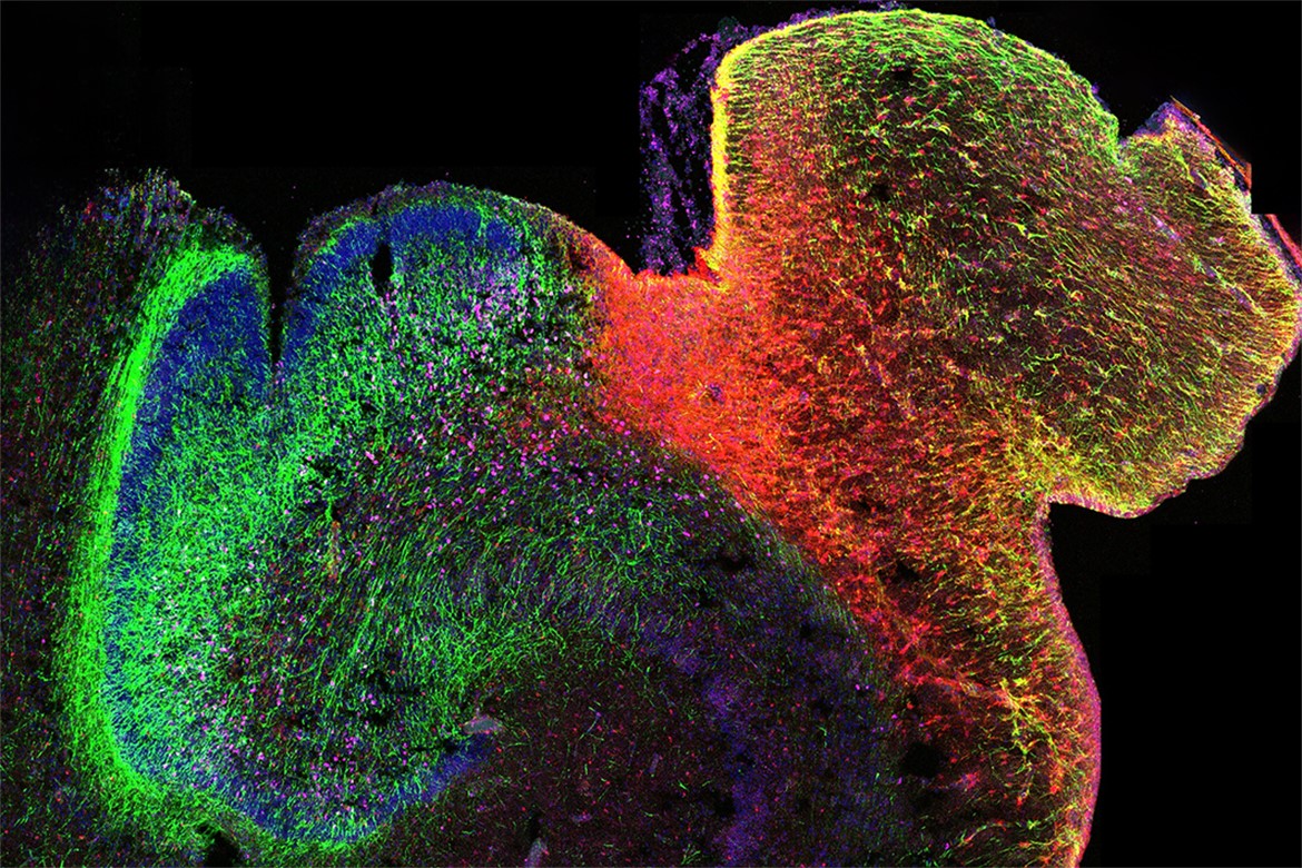
The study was a collaboration between scientists at the Salk Institute, UC Los Angeles, UC San Francisco, UC San Diego, and Seoul National University. The team created the first map of DNA modification in the hippocampus and prefrontal cortex—two regions of the brain critical to learning, memory, and emotional regulation. These areas are also frequently involved in disorders like autism and schizophrenia.
The findings were published in Nature on October 9, 2024.
The researchers hope the data resource, which they’ve made publicly available through an online platform, will be a valuable tool scientists can use to connect genetic variants associated with these conditions to the genes, cells, and developmental periods that are most sensitive to their effects.
“Growing a healthy human brain is a tremendous feat,” says study co-author Joseph Ecker, Salk professor, Salk International Council Chair in Genetics, and Howard Hughes Medical Institute investigator. “Our study establishes an important database that captures key epigenetic changes that occur during brain development, in turn bringing us closer to understanding where and when failures arise in this development that can lead to neurodevelopmental disorders like autism.”
To produce the map, the research team used a cutting-edge sequencing approach called single nucleus methyl-seq and chromatin conformation capture, or snm3C-seq. This technique enables researchers to simultaneously analyze two epigenetic mechanisms that control gene expression on a single-cell basis: chemical changes to DNA known as methylation and chromatin conformation, the 3D structure of how chromosomes are tightly folded to fit into nuclei.
Figuring out how these two regulatory elements act on genes that affect development is a critical step to understanding how errors in this process lead to neuropsychiatric conditions.
“The vast majority of disease-causing variants we’ve identified are located between genes on the chromosome, so it’s challenging to know which genes they regulate,” says study co-lead author Chongyuan Luo, an assistant professor of human genetics at the David Geffen School of Medicine at UCLA. “By studying how DNA is folded inside of individual cells, we can see where genetic variants connect with certain genes, which can help us pinpoint the cell types and developmental periods most vulnerable to these conditions.”
For example, autism spectrum disorder is commonly diagnosed in children aged 2 and over. However, if researchers can gain a better understanding of the genetic risk of autism and how it impacts development, they can potentially develop intervention strategies to help alleviate the symptoms of autism, like communication challenges, while the brain is developing.
The research team analyzed more than 53,000 brain cells from donors spanning mid-gestation to adulthood, revealing significant changes in gene regulation during critical developmental windows. In capturing such a broad spectrum of developmental phases, the researchers were able to assemble a remarkably comprehensive picture of the massive genetic rewiring that occurs during critical timepoints in human brain development.
One of the most dynamic periods comes around the midpoint of pregnancy. At this time, neural stem cells called radial glia, which have produced billions of neurons during the first and second trimesters, stop producing neurons and begin generating glial cells, which support and protect neurons. At the same time, the newly formed neurons mature, gaining the characteristics they need to fulfill specific functions and forming the synaptic connections that enable them to communicate.
This stage of development was overlooked in previous studies, the researchers say, due to the limited availability of brain tissue from this period.
“Our study tackles the complex relationship between DNA organization and gene expression in developing human brains at ages typically not interrogated: the third trimester and infancy,” says study co-lead author Mercedes Paredes, an associate professor of neurology at UCSF. “The connections we’ve identified across different cell types through this work could untangle the current challenges in identifying meaningful genetic risk factors for neurodevelopmental and neuropsychiatric conditions.”
The findings also have implications for improving stem cell-based models, such as brain organoids, which are used to study brain development and diseases. The new map offers a benchmark for scientists to ensure these models accurately replicate human brain development.
“Neuropsychiatric disorders, even those emerging in adulthood, often stem from genetic factors disrupting early brain development,” says Luo, who previously trained in Ecker’s lab at Salk. “Our map offers a baseline to compare against genetic studies of disease-affected brains and pinpoint when and where molecular changes occur.”
The group’s efforts are part of the National Institutes of Health’s BRAIN Initiative Cell Atlas Network, or BICAN, which aims to build reference brain cell atlases that will provide a foundational framework for studying brain function and disorders.
Other authors include Jingtian Zhou and Jesse Dixon of Salk; Matthew Heffel, Yi Zhang, Kangcheng Hou, Kevin Abuhanna, Joseph Galasso, Terence Li, Eleazar Eskin, and Bogdan Pasaniuc of UC Los Angeles; Dong-Sung Lee of Seoul National University; Oier Pastor Alonso, Ryan Ziffra, and Tomasz Nowakowski of UC San Francisco; Colin Kern, Chu-Yi Tai, Carlos Garcia Padilla, Mahsa Nafisi, Yi Zhou, Eran Mukamel, Quan Zhu, and Bogdam Bintu of UC San Diego; Anthony Schmitt of Arima Genomics; Maximilian Haeussler and Brittney Wick of UC Santa Cruz; Martin Jinye Zhang of Carnegie Mellon University and the Harvard T.H. Chan School of Public Health; and Fangming Xie of UC Los Angeles and UC San Diego.
The work was supported by the National Institute of Mental Health, National Human Genome Research Institute, National Institute on Drug Abuse, Simons Foundation, Roberta and Oscar Gregory Endowment in Stroke and Brain Research, Chan Zuckerberg Biohub, National Research Foundation of Korea, Shurl and Kay Curci Foundation, and California Institute for Regenerative Medicine.
DOI: 10.1038/s41586-024-08030-7
JOURNAL
Nature
AUTHORS
Matthew G. Heffel, Jingtian Zhou, Yi Zhang, Dong-Sung Lee, Kangcheng Hou, Oier Pastor Alonso, Kevin D. Abuhanna, Joseph Galasso, Colin Kern, Chu-Yi Tai, Carlos Garcia Padilla, Mahsa Nafisi, Yi Zhou, Anthony D. Schmitt, Terence Li, Maximilian Haeussler, Brittney Wick, Martin Jinye Zhang, Fangming Xie, Ryan S. Ziffra, Eran A. Mukamel, Eleazar Eskin, Tomasz J. Nowakowski, Jesse R. Dixon, Bogdan Pasaniuc, Joseph R. Ecker, Quan Zhu, Bogdan Bintu, Mercedes F. Paredes, Chongyuan Luo
Office of Communications
Tel: (858) 453-4100
press@salk.edu
The Salk Institute is an independent, nonprofit research institute founded in 1960 by Jonas Salk, developer of the first safe and effective polio vaccine. The Institute’s mission is to drive foundational, collaborative, risk-taking research that addresses society’s most pressing challenges, including cancer, Alzheimer's disease, and agricultural vulnerability. This foundational science underpins all translational efforts, generating insights that enable new medicines and innovations worldwide.