November 30, 2021
Salk scientists have discovered that neurons deep in the brain’s cortex are the first to compute which side of a visual border is an object and which side is background
Salk scientists have discovered that neurons deep in the brain’s cortex are the first to compute which side of a visual border is an object and which side is background
LA JOLLA—In the classic “Rubin’s vase” optical illusion, you can see either an elaborate, curvy vase or two faces, noses nearly touching. At any given moment, which scene you perceive depends on whether your brain is viewing the central vase shape to be the foreground or background of the picture.

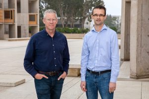
Now, Professor John Reynolds and Senior Postdoctoral Fellow Tom Franken have made headway into understanding how the brain decides which side of a visual border is a foreground object and which is background. The research, published on November 30, 2021 in the journal eLife, sheds light on how areas of the brain communicate to interpret sensory information and build a picture of the world around us.
“The way that the brain organizes and generates a representation of the outside world is still one of the biggest unknowns in neuroscience today,” says Reynolds, holder of the Fiona and Sanjay Jha Chair in Neuroscience. “Our research provides important insights into how the brain processes borders, which could lead to a better understanding of psychiatric conditions where perception is disrupted, such as in schizophrenia.”
When you view a scene in front of you, individual neurons in the brain’s cortex each receive information about a minuscule region of the scene. Neurons receiving information from the border of an object thus have little initial context about which side is foreground. However, scientists previously discovered a set of cells that very quickly signal which side of the border belongs to the object (“ border ownership”); after all, depth perception and the ability to pick out objects in front of you is critical to survival—is that a curb or a shadow, a rock or a cave?
Exactly how these neurons in the brain compute border ownership has been unclear. Some scientists hypothesized that as information from the eye passes through the brain, into successively more downstream (deeper) areas, additional computations occur in each area until your brain builds a model of the visual scene. This is called the “feedforward” pathway. But other scientists hypothesized the importance of the “feedback” pathway, in which downstream areas of the brain must first process information, and then send these clues back to neurons in upstream areas, to help them figure out border ownership.
Reynolds and Franken set out to determine which hypothesis was correct. They used electrodes to record the activity of neurons in different layers of the brain’s cortex as animals viewed an image of a square object on an otherwise blank background. The scientists first determined which particular neurons were processing information from a small part of the border that demarcates the square and the background; then they measured the timing of border ownership signals in these neurons and compared this for neurons in different layers.

“What we found is that the earliest signals on border ownership occur in neurons in the deep layers of the brain’s cortex,” says Franken, who is a physician-scientist and supported by a K99 Pathway to Independence Award from the National Institutes of Health. “This supports the importance of the feedback pathway for deciphering borders, because feedback connections arrive at and leave from neurons in deep layers.”
The researchers also observed that neurons stacked vertically in different layers in the cortex tended to share the same preference of border ownership. For example, certain columns of neurons preferred scenes where the left side of a border was the object, while other columns of neurons preferred scenes where the right side of a border was the object. Franken explains that these findings suggest that feedback might actually be organized in a systematic way, a promising avenue for further research.
“As we come to understand the architecture of the brain and how ensembles of neurons communicate with each other to build up our internal representation of the external world, we are better positioned to develop diagnostic tools and treatments for brain disorders in which these internal representations are distorted, such as schizophrenia,” says Franken. “The hallucinations and delusions associated with schizophrenia may be associated with the disruptions of feedforward-feedback loops.”
Next, Franken will follow up on these results with experiments to investigate how information conveyed by feedback contributes to the processing of borders.
The work was supported by grants from the George E. Hewitt Foundation for Medical Research, a NARSAD Young Investigator Grant from the Brain & Behavior Research Foundation and the National Eye Institute of the National Institutes of Health.
DOI: https://doi.org/10.7554/eLife.72573
JOURNAL
eLife
AUTHORS
Tom Franken and John Reynolds
Office of Communications
Tel: (858) 453-4100
press@salk.edu
Unlocking the secrets of life itself is the driving force behind the Salk Institute. Our team of world-class, award-winning scientists pushes the boundaries of knowledge in areas such as neuroscience, cancer research, aging, immunobiology, plant biology, computational biology and more. Founded by Jonas Salk, developer of the first safe and effective polio vaccine, the Institute is an independent, nonprofit research organization and architectural landmark: small by choice, intimate by nature, and fearless in the face of any challenge.